Individuals
249
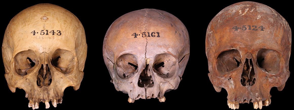
Three crania from the Green Ground
249
97
50
8
84
The Green Ground was a post-medieval graveyard on Portugal Street used by the Parish of St Clement Danes (western central London) from 1638.
It was notorious for its overcrowded and unsanitary conditions.
The graveyard was closed around 1850 to make way for an extension to the old King's College Hospital, now the Library of the London School of Economics.
The Green Ground graveyard occupied about one third of an acre of land on Portugal Street. It was predominantly a burial place for the poor or destitute and was notorious for its overcrowded and unsanitary conditions.
The Green Ground was closed around 1850 following a major cholera epidemic and the subsequent passing of a number of Burial Acts by Parliament. The majority of inner London graveyards were closed at this time.
The land was cleared in 1852 to make way for an extension to King's College Hospital and a number of recovered crania and mandibles were presented to the Royal College of Surgeons.
After the College was bombed in 1941, the remains that survived were transferred to the Natural History Museum.
Given the high number of dental and other diseases, it is probable that these bones were collected specifically for the study of these pathologies or for use as teaching specimens.
Cranium of an adult, probable male, with metopic suture. There is a healed depression fracture approximately seven millimetres in diameter on the left frontal eminence, caused by blunt force trauma.
There is slight porosity on the vault and moderate porosity on the hard palate and sphenoid, suggestive of poor health or metabolic disease.
On the teeth there is one carious lesion, slight recession of the alveolar bone and dental calculus, indicative of poor dental hygiene. Iron staining can be seen on the left and right sides of the frontal bone and on the occipital bone, probably resulting from contact with coffin nails.

Cranium of adult male with arrow pointing to healed depression fracture
Cranium of an adult of indeterminate (ambiguous) sex. The cranium was dissected during curation and due to a lack of suture fusion the bones of the skull are disarticulated. This individual had syphilis.
There are numerous foci of caries sicca (lesions) on the cranial vault and sclerotic stellate scars, giving it a 'moth-eaten' appearance.
These lesions are limited to the destruction of the outer table and have not penetrated the inner table. The inner surface of the cranium has increased vascularity and porosity is present on the occipitals and within the orbits, suggestive of further poor health.
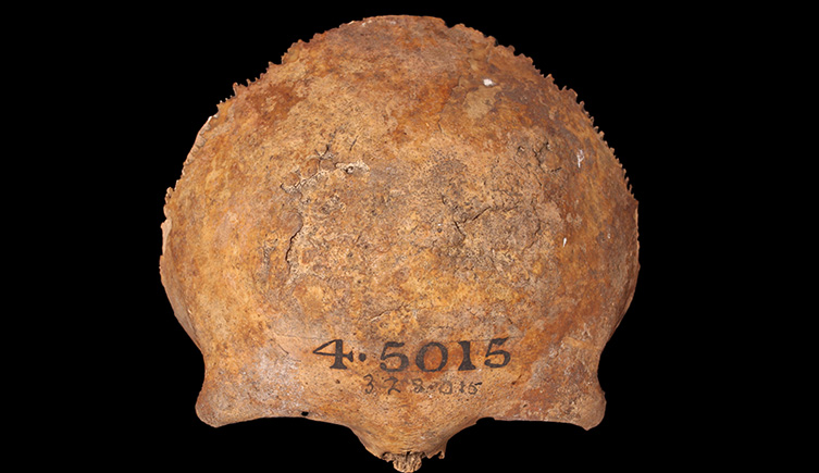
The frontal bone of an adult with syphilis
Cranium of a young adult male. The left nasal bone was transversely fractured ante-mortem (during life) and since healed. There are three benign bony overgrowths, or button osteomas, on the cranium.
There is slight porosity on the parietal and frontal bones, and linear enamel hypoplasia on the left upper second premolar, indicative of poor diet and health.The individual also had dental calculus and two carious lesions on the teeth.
There is one small copper stain and two small iron stains on the cranium, probably from contact with a copper shroud pin and coffin nails.
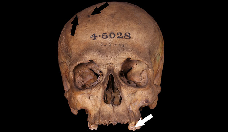
Cranium of a young adult male. White arrow points to interdental carious lesion, black arrows to two small osteomas.
Cranium of a probable young adult male. There is a small rounded depression fracture on the right frontal eminence caused by blunt force trauma.
This individual suffered from poor health, evident from porosity throughout the cranium and the presence of linear enamel hypoplasias on at least two teeth.
There are small amounts of desiccated skin adherent.

Cranium of a probable young adult male. Arrow points to rounded depression fracture. Desiccated skin can be seen on the left maxilla.
Cranium of an adult male. This individual presents a case of cribra orbitalia as coalesced porosity is observed in both orbits.
There is also porosity at the bregma, on the palate and at the base of greater wing of sphenoid.
These suggest that this individual suffered from poor nutrition or metabolic disease.
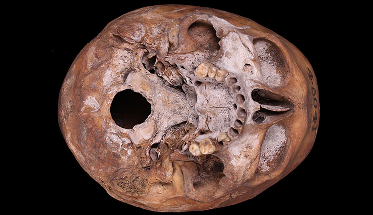
Cranium of an adult male with cribra orbitalia
Cranium of an adult female with more dental wear than is commonly seen for the post-medieval period.
This could have been due to a course diet or a work-related activity that caused abrasion to individual's teeth.
There is also complete destruction of the second right molar crown due to caries (tooth decay) and linear enamel hypoplasia, suggestive of poor dental hygiene and poor health.
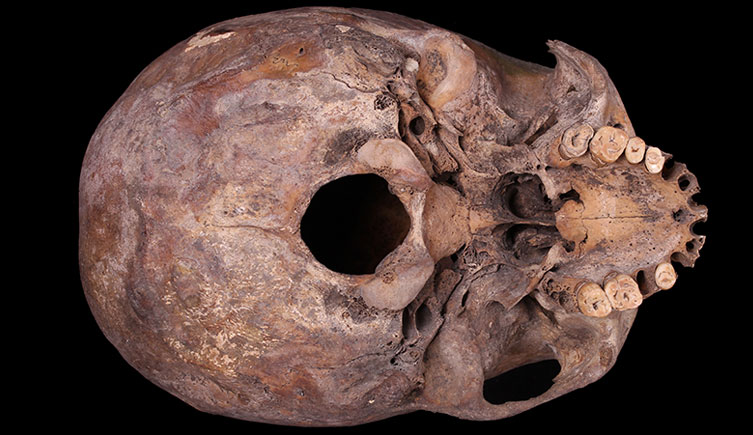
Cranium of an adult female with relatively advanced dental wear and destruction of right second molar crown due to caries
Cranium of an adult, probable female. On the basal cranium there are two developmental anomalies. The left occipital condyle has two articular facets and is longer that the right, and a distinct precondylar tubercle, a bony exostosisis, is present at the anterior midline of the foramen magnum margin.
This individual had arthritis of the right temporomandibular joint, evident from bone loss, porosity and mild lipping to the joint surface's lateral-anterior border, as well as perforation (with sclerotic reaction) and an enthesophyte (ossified tissue) on the tympanic part of the right temporal. All teeth were lost ante-mortem (during life), and the alveolar bone subsequently resorbed. A large facially draining abscess is present at the rear of the right maxilla, with clear evidence of healing on the margins of the perforation.
Cranium of an adult male. This individual suffered from a sharp and blunt force trauma to the left side of the face above the orbit. This caused an oval depression fracture as well as a V-shaped depression which at its apex perforated the cranium. It is probable that this caused additional fracturing and shape deformation of the left orbit, although some further fractures occurred post-mortem. The trauma was not the immediate cause of death as there is evidence of healing to the bone, as well as stellate scars suggesting that there was an infection associated with the trauma, but the eventual cause of death is unknown.
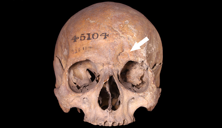
Cranium of an adult with arrow pointing to blunt and sharp force trauma
Cranium and mandible of an adult male. This individual smoked a pipe, which has caused the left upper second incisor and left upper canine to be worn down into a pipe facet. Smoking probably caused the molars on the left side have a large amount calculus on the occlusal (chewing) and facial aspects, caries and staining, perhaps also exacerbated by poor dental hygiene. This individual also suffered from poor health when they were young, as many of the teeth have linear enamel hypoplasias.

Skull of an adult male with arrow pointing to pipe facet
Cranium of an adult male. There is distinct bunning (protrusion) of the occipital bone due to a number of wormian bones in the lambdoidal suture. Wormian bones are abnormal bones that occur within the sutures of the cranium. They are fairly common, particularly in the lambdoidal suture, but the number seen in this case is unusual.
The London human remains collection has been digitised
If you would like to use any specimens for research, please get in touch
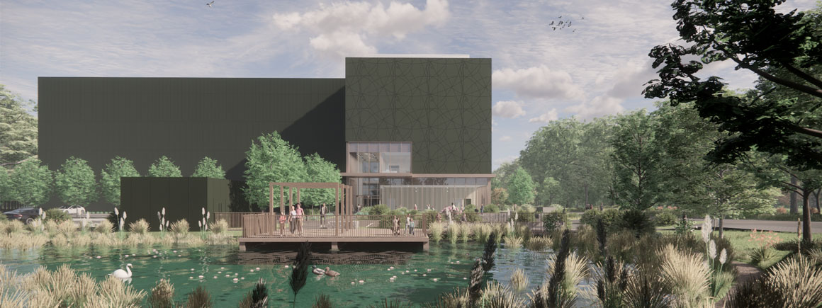
Access to some collections will be affected as we prepare for the move to our new collections, science and digitisation centre.
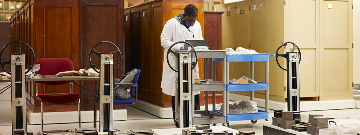
Scientists and collections management specialists can visit the collections and borrow specimens for research.
Our duty is to provide a safe and secure environment for all of our collections.