Mineral sample laboratory
The museum’s mineral preparation laboratory prepares specimens from the collections for further analysis with SEM, geochemical and optical description/identification.

The museum’s mineral preparation laboratory prepares specimens from the collections for further analysis with SEM, geochemical and optical description/identification.
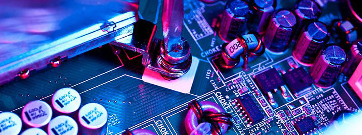
We repair and modify specialist electronic equipment.
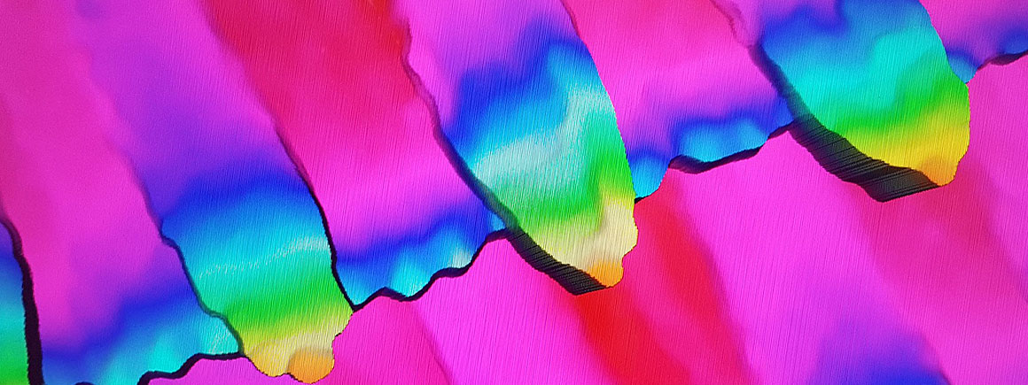
The 3D visualisation laboratory has three scanners for digitising large-scale objects and specimens sized from 2cm to 3m.
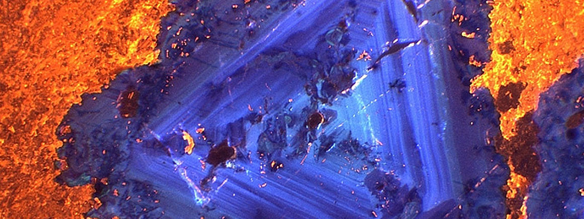
Our electron probe microanalysis instruments allow us to conduct qualitative and quantitative elemental analysis of samples.
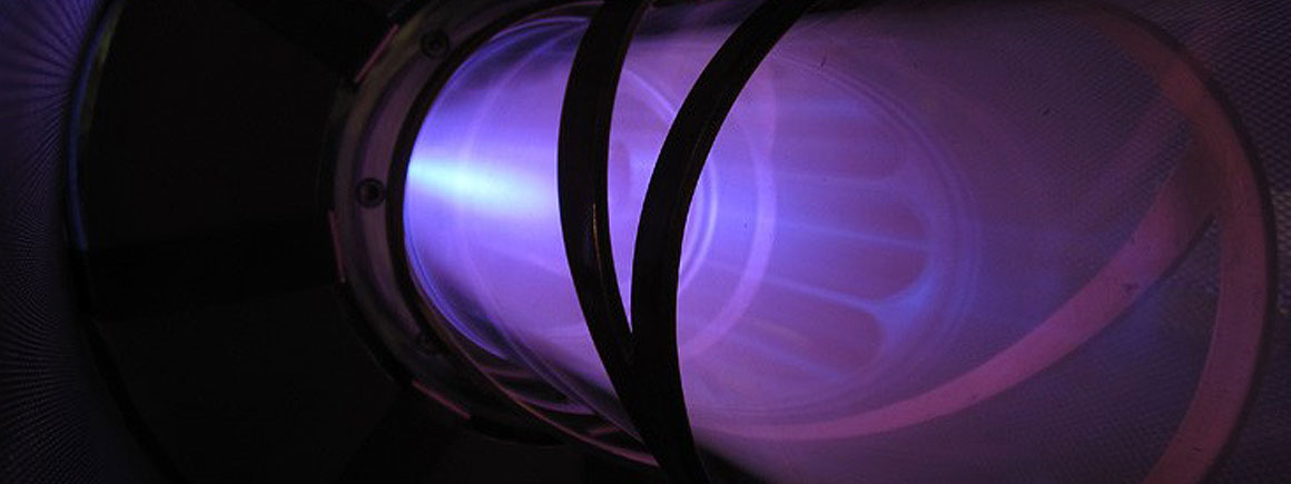
We use specialist techniques to determine the concentrations of elements and chemical species in a wide range of samples.
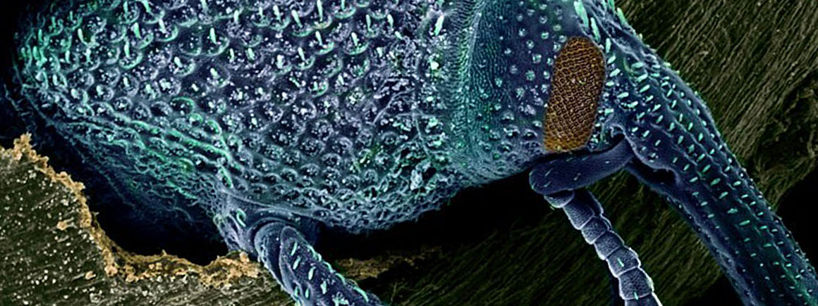
The electron microscope is a type of microscope that uses a beam of electrons to create an image of the specimen.
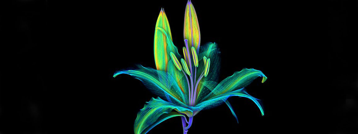
Micro-CT is a non-invasive and non-destructive technique that uses X-rays to create 3D models of the internal and external features of specimens.
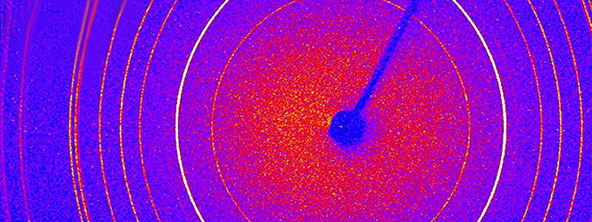
A tool for phase identification, quantification and crystal structure analysis.
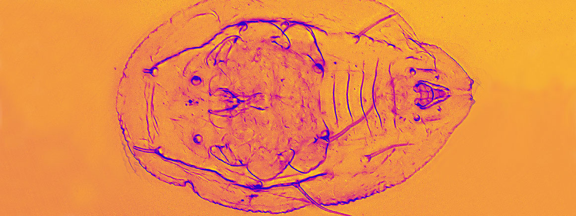
The Light Microscopy Facility houses state-of-the-art microscopy systems that allow scientists from all over the world to see beyond the limits of the naked eye.
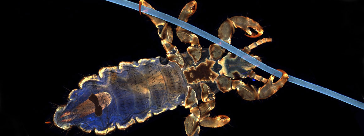
In confocal microscopy, an object is scanned using a laser beam to build up the image a line at a time.